
A team of health technology researchers has unveiled a rapid, non-invasive method for detecting early signs of impaired circulation in diabetic feet. The “foot perfusion scan”—a near-infrared fluorescence-based test—takes only 60 seconds and promises to identify early microvascular issues before they escalate, potentially preventing many amputations. Its speed and simplicity could make it a game-changer in diabetic foot screening and early intervention.
Early detection of peripheral arterial disease (PAD) in diabetic individuals has long challenged clinicians. Traditional tools like the ankle–brachial pressure index (ABPI) and toe–brachial indices can be unreliable in this group due to calcified arteries and neuropathy, and more advanced tests like transcutaneous oxygen or Doppler studies often require significant time, cost, and expertise. The need for a fast, accurate, and accessible alternative has thus been a long-sought goal in diabetes management.
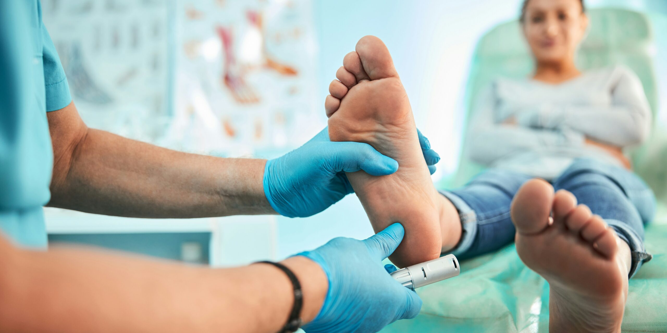
The foot perfusion scan works by injecting indocyanine green (ICG), a fluorescent dye, and then using near-infrared imaging to visualize blood flow beneath the skin. As the dye travels through the microvasculature, a camera captures real-time flow dynamics—revealing perfusion delays or poor circulation. This method is far quicker than conventional assessments and can assess multiple regions simultaneously .
In recent trials, researchers reported that ICG near-infrared imaging uncovers subtle variations in foot perfusion among diabetic patients—even those without visible ulcers or claudication. Data show areas with delayed or reduced dye uptake often correspond to zones at high risk for ulceration or deteriorating tissue health. One study found that even in the presence of stiff arteries and misleading physical symptoms (like warm, pink feet), optical perfusion imaging accurately flagged underlying ischemia.
The implications of such early detection are vast. Diabetic foot ulcers affect roughly 6–13% of patients, depending on region, and carry a high rate of non-healing and progression to amputation . Mortality after foot ulceration can be greater than 50% over five years—comparable to many aggressive cancers. By identifying circulatory deficits before ulcers form, care teams can intervene earlier with shoe off-loading, vascular referrals, or lifestyle adjustments, significantly reducing risk.
Clinicians who used the scan report that results, available within a minute, were immediately actionable. Dr. Sarah Patel, one of the lead researchers, stated: “This test allows us to see microcirculation in real time. When we detect poor perfusion, we can swiftly adjust treatment—often before the patient even has symptoms.” By empowering providers with on-the-spot data, the technology streamlines triage and improves patient understanding of their health status.
Besides its clinical merits, the scan’s simplicity could facilitate broader community and even home use. It requires only topical dye and a compact imaging device. With minimal training, general practitioners, podiatrists, or nurse practitioners can administer it during routine visits, dramatically expanding screening capacity beyond specialized vascular labs.
Still, experts caution that perfusion scans should complement—not replace—comprehensive clinical assessments. Factors such as infection, edema, or recent injury can alter perfusion readings. As one commentary noted, “functional testing offers powerful signals, but must be integrated with patient history, bloodwork, and risk profiling” . Ongoing trials are now comparing the scan’s predictive accuracy to standard measures like toe pressures or transcutaneous oxygen flux.
Major next steps include multicenter validation involving diverse patient populations—especially in primary care settings—and cost-benefit analyses to establish reimbursement models. Early economic modeling, based on amputation reduction alone, suggests a single scan costing $200 could save healthcare systems tens of thousands per averted limb loss.
If adopted widely, the 1-minute scan could dramatically shift standard clinical workflows. Rather than awaiting ulceration or overt PAD symptoms, a perfusion scan during a simple foot exam might become the norm—prompting timely vascular referrals, tailored footwear, and optimized glycemic or blood pressure control.
In a broader context, the technology exemplifies the growing potential of optical imaging in medicine. Similar non-invasive methods are under development for wound healing, peripheral vascular assessment, and even evaluating flap viability in reconstructive surgery .
In summary, the foot perfusion scan offers a rapid, precise, and scalable tool for early PAD detection in diabetics—potentially preventing ulcer formation and drastically reducing amputation rates. Its ease and immediacy could make it a routine part of diabetic foot exams, delivering timely care and long-term cost savings.
As a next step, large-scale clinical trials and health economic studies will determine how quickly this innovation transitions from research labs to everyday practice. For millions of people living with diabetes, a one-minute scan may soon make the difference between limb preservation and life-altering amputation.